Under the microscope
Established in 2015, the Mackay School of Earth Sciences and Engineering Microbeam Laboratory houses various state-of-the-art instruments for use by researchers at the University of Nevada, Reno and beyond.
Learn about available instruments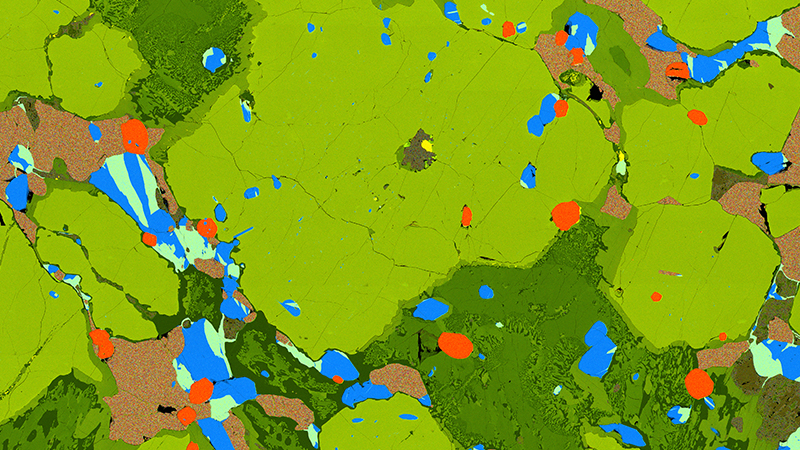
Recent news from the Mackay School

Workshop explores a broader vision for lithium: fusion and fission energy applications
The interdisciplinary workshop brought scientists, businesspeople and government officials to the University campus

Richard Goldfarb reflects on his education at the University
The alumnus has travelled widely in his economic geology career

Interdisciplinary conference explores branching structures
The conference was held at the University of Nevada, Reno at Lake Tahoe in memory of late math professor