Director and staff
Core director
- Lars Strother, Ph.D.
- Email: lars@unr.edu
- Phone: 775-384-7109
Research manager
- Sean O'Neil, Assistant Director
- Email: seano@unr.edu
This facility is designed to support usage and analysis of data from a variety of structural and functional neural imaging technologies and techniques: magnetic resonance imaging (MRI), high-density array electroencephalography (HD EEG) and near-infrared spectrospcopy (NIRS). The MRI facility is housed at Renown Health and consists of two 3T scanners (one Philips Ingenia, and one Siemens Skyra). A neuroimaging partnership with Renown Health has been in place since 2014. The newer scanner (Siemens Skyra) is currently being equipped to improve capabilities and access for University researchers. Two HD EEG systems (Electrical Geodesics and Biosemi) the NIRS system (TechEn) are located on campus (MSS 3rd floor). The Neuroimaging Core’s technologies are used by multiple labs within neuroscience and related departments. All equipment sees frequent use throughout the year.
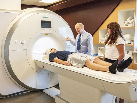
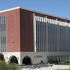
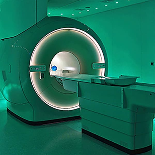
Magnetic Resonance Imaging (MRI) is a non-invasive technology used by researchers to study brain anatomy and function. MRI uses a strong magnetic field and radio waves to measure signals emitted by excited hydrogen atoms present in various types of bodily tissue. These signals are converted into images that show different degrees of contrast for different tissue types, which allows researchers to create detailed anatomical images of the brain. Functional MRI (or fMRI) uses MRI technology to measure brain activity, which can then be superimposed on images of brain anatomy from MRI. Unlike MRI, fMRI measures blood-oxygen-level dependent (BOLD) signals that indicate the amount of blood in different areas of the brain. Blood volume increases as a function of neural activity because the brain does not store the glucose needed to provide energy to the metabolically "expensive" activity of its neurons, and blood must be shunted to brain regions that are most active. Like EEG, fMRI measures the activity of large populations of neurons. However, the spatial resolution of fMRI is much greater than that of EEG-fMRI has fine spatial resolution but poor temporal resolution (unlike EEG). Researchers at the University of Nevada, Reno, are using fMRI to study a broad spectrum of cognitive and perceptual abilities including vision, memory, and uniquely human skills such as reading.
MRI Prescreening Form - Complete prior to visit
Directions to MRI (Google Maps)
The MRI facility is located in the Renown South Meadows building at 10085 Double R Blvd. It is located east of the emergency room, past the DaVita Dialysis center. (Do not enter the DaVita Dialysis center.) Enter the exterior door and the MRI facility is to your right.
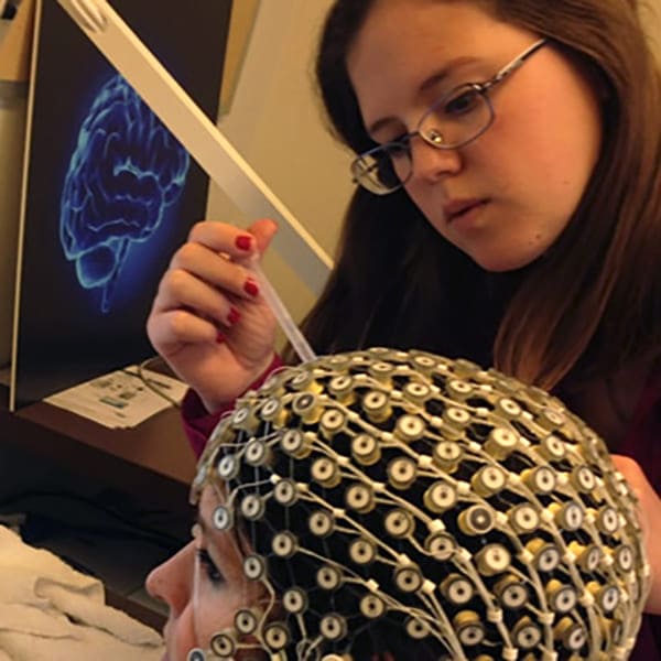
Electroencephalography (EEG) is a non-invasive technology that measures voltage fluctuations on the scalp that result from the synchronized activity of large groups of neurons in the brain. In some cases, this activity is associated with a particular stimulus or task. For instance, evoked potentials (EPs) and event-related potentials (ERPs) involve the time-locking of stimulus presentation (e.g. visual or auditory) to the measurement of scalp potentials. EEG has high temporal resolution (on the order of milliseconds), which gives it an advantage over other neuroimaging technologies such as fMRI and fNIRS. Because of its high temporal resolution, EEG activity can be correlated with specific stimulus events and behaviors at a very fine time scale. On the other hand, the spatial resolution of EEG is limited. To overcome this limitation, researchers at the University of Nevada, Reno use high-density array EEG systems. Researchers may use either a 256-channel EGI Net Amps system or a 128-channel Biosemi ActiveTwo system. Each system has its own strengths and can be used to measure brain activity including determining the source of EEG signals in the brain. State of the art neuroimaging software is available for processing and analysis.
EGI EEG system calendar Effie Mona Mack, room 302
Biosemi EEG system calendar Effie Mona Mack, room 310
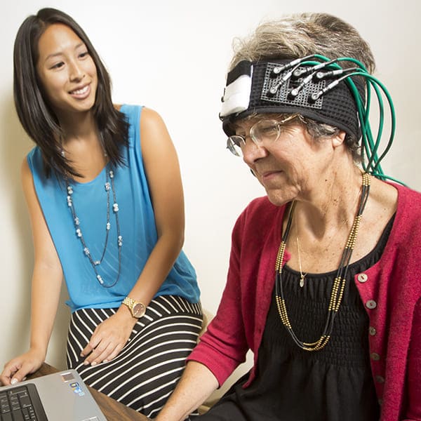
Near-infrared spectroscopy (NIRS) is a non-invasive means of measuring brain activity. If you hold a flashlight to the palm of your hand in the dark you can see that its light will travel through several centimeters of tissue. NIRS affords optical brain imaging at centimeter depth by projecting near-infrared wavelength light through the skull and measuring how much has been absorbed by the hemoglobin in your blood. That is, NIRS indirectly measures brain activity by measuring the amount of oxygenated and deoxygenated hemoglobin in a particular region of the brain-this relies on the fact that active areas of the brain require blood to sustain neural activity, which is metabolically taxing. NIRS thus allows researchers to "see" brain activity, which is essential to understanding the function of the human brain. We therefore sometimes refer to this use of NIRS technology as "functional NIRS" or fNIRS. Researchers at the University of Nevada, Reno, are using fNIRS and other neuroimaging technologies-including functional MRI (fMRI) and high-density array EEG to study a variety of human cognitive and perceptual abilities.
Mack Social Science, room 305
This MRI Facility houses a new Siemens 32-channel 3T Skyra (Siemens Healthcare, Germany) and a Philips 32-channel 3T Ingenia (Philips Medical Systems, The Netherlands). Both scanners have wide bores and are equipped with MR-compatible technologies for conducting experiments with human subjects. The Philips system has been used by up to ten labs over the course of five years; it will be phased out later this year and the Siemens will be used for most if not all current and future scanning. In addition to high resolution images of brain anatomy (structural MRI or sMRI), both scanners support functional MRI (fMRI) and diffusion imaging of long-range white matter tracts, and thus adequately support all UNR neuroimaging needs.
"Functional" MRI (fMRI) differs from high-field strength "structural" MRI (sMRI) by its ability to image beyond anatomical detail into the functional workings of the brain, how the brain thinks, and where it all takes place. The specific type of fMRI being utilized in these research projects is known as "BOLD" fMRI, more scientifically known as "neurovascular coupling". As important and exciting as all of this is, there are also other techniques that fall under the general heading of fMRI as well.
Fractional anisotropy (FA), featuring directionally color-coded fiber tracts, can map out deep white matter axonal pathways which connect all of the eloquent sites in the brain identified with the BOLD fMRI. The FA maps can easily be converted into Diffusion Tensor Images (DTI) in which the exquisite neuroanatomy of the connections in the brain are displayed and studied.
MR Spectroscopy (MRS) and Perfusion MRI (pMRI) also fit under the heading of fMRI. MRS deals with in vivo examination of hydrogen spectra in selected regions of brain, while pMRI allows assessment of the amount of blood flowing into and through any portion of brain anatomy.
The Siemens 32-channel 3T Skyra system is fully equipped (as of Fall 2019) for advanced neuroimaging, and in many ways surpasses the capabilities of the Philips Ingenia system. This Siemens system has a fast gradient system that provides high-speed spatial encoding, a 64-channel data acquisition system with digital wireless technology to improve SNR and temporal stability, and a dual-channel RF transmitter system for reduction of dielectric effects, and more flexible RF pulse design. The gradient rise time (200 mT/m/ms), peak gradient strength (45 mT/m per axis), and duty cycle (100% using full gradient strength on all three axes) are the highest specifications in the industry for whole-body systems. Also, the gradients have a balanced geometric design that results in less acoustic noise generation. Every hardware and software option that Siemens offers for neuroimaging is installed on this MRI system. Parallel imaging capabilities in one and two-dimensions enable EPI acquisitions at higher temporal resolution and with less geometric distortion. In addition, a Master Research Agreement with Siemens Healthcare makes available advanced "works-in-progress" pulse sequences for EPI and structural neuroimaging to deal with specific technical challenges.
The Siemens system is equipped with fMRI-presentation hardware, screen and response pads. It is also equipped with a vPixx eye-tracking system. The TRACKPixx3 MRI/MEG is a 2 kHz eye/gaze-tracking solution compatible with MRI and MEG environments. The TRACKPixx3 is versatile, supporting both monocular and binocular tracking with a single mechanical configuration. The TRACKPixx3 does not require a dedicated PC to process eye images and generate gaze information; all image processing is performed within the TRACKPixx3 hardware. Gaze data can be logged within the TRACKPixx3 and retrieved by the testing PC with a simple low-latency USB interface. The TRACKPixx3 video feed can be accessed directly through a console display for real-time visualization and adjustment of the tracker. A scene camera can be connected to the tracker to monitor the experiment. These video feeds can also be accessed through the USB interface for remote control of the TRACKPixx3.
The Philips Ingenia 3.0T system comes with a dStream digital broadband architecture and a channel independent RF technology, which results in an up to 40% more SNR. dStream digitizes the signal right in the coil, eliminating noise influences typical of analog pathways, to capture the MR signal without predistortion or compression. A fiber-optic connection from the coil to the image reconstructor enables lossless broadband data transmission. This Ingenia 3.0T system has a higher order shim function which offers advanced shimming capabilities to obtain improved image quality in field-sensitive applications and techniques such as single-voxel spectroscopy, chemical shift imaging, single-shot EPI and balanced FFE. This Ingenia 3.0T system features high performance whole body, non-resonant, self-shielded gradient technology with new amplifiers that deliver high peak and slew rates for the demanding requirements of the latest and emerging clinical imaging techniques. The Quasar Dual gradient system provides industry leading performance specifications for peak strength and slew rate with a dual mode capability that optimizes advanced applications requiring very high peak mode capabilities. The maximum gradient amplitudes and slew rates corresponding to the dual mode are 80 mT/m, 100 mT/m/ms and 40 mT/m, 200 mT/m/ms respectively. This Ingenia 3.0T system has a multiple RF sources, which adapts the RF signals to suit each individual patient. This results in a faster scan, enhanced image uniformity/consistency, over a broader range of applications. This Ingenia 3.0T system features MultiBand SENSE which allows you to use state-of-the-art acceleration factors in the brain by simultaneously exciting multiple slices. Due to a shorter minimum TR for fMRI, larger anatomical coverage or higher temporal resolution can be used. In the DWI/DTI sequences larger anatomical coverage or higher number of diffusion directions can be acquired. With MultiBand SENSE, fMRI and DTI exams can be performed with high speed and high resolution, simultaneously.
This Ingenia 3.0T system is equipped with a Multi-nuclear spectroscopy (MNS) system, which provide the ability to perform 13C, 31P, 7Li, 23Na, 19F and other nuclei spectroscopy and imaging. The multiple RF amplifiers in this system includes two 18 kW solid-state 1H channel narrowband amplifier and one 4 kW broadband (10-130 MHz) Multi-nuclear amplifier.
This Ingenia 3.0T system has a bore diameter of 60 cm and provides a full-size 50 cm field-of-view. Analog to digital signal conversion on the coil, 70cm bore, multi-transmit, Omega HP Gradients (slew rate 200 mT/m/ms, maximal gradient strength 45 mT/m), unlimited RF channels, fMRI-presentation hardware, screen and response pads.
Our Neuroimaging facilities in MSS 300 feature 2 high density EEG systems. They work in a similar fashion to “traditional” EEG in that they measure microvoltage electricity related to superficial neural activity, but our systems cover a much larger area of the cortex, allowing for more simultaneous data collection and more complex analyses that require whole-head data. Although the number of channels is greatly increased, setup time is not burdensome as individual site locations don’t need to be measured and scalp preparation is minimal. Our two systems differ in a few key details, but are both suitable for a diverse number of projects. They are each used by several labs and dozens of individual users, and in use for numerous techniques including traditional neural wave-based analyses, event-related potentials, steady-state visual evoked potentials, and time-frequency analysis.
The Electrical Geodesics Inc./Philips system (EGI; Eugene, Oregon) features 256-channel HydroCel caps recording at 1000 Hz. This system allows for easy setup of routine EEG in human participants using an electrolyte solution-based interface. EEG data can be corresponded with MRI data using the Geodesic Photogrammetry System, improving spatial identification of neural sources. The Biosemi ActiView system (Amsterdam, Netherlands) records 128 channels at 2048 Hz and uses a gel-based interface for recording. It features active electrodes which allow for amplification nearer to the source and corresponding high signal to noise ratio. Optional skin-contact electrodes can be used for artifact detection, referencing, or electromyography. The Biosemi also uses a 1080p LCD monitor (Cambridge Research Systems Display++) which allows for research suitable color and timing reproduction, and additionally removes the need to calibrate the monitor, normally a time-consuming process. This system is portable and battery-powered, intended to isolate the participant from mains power and decrease 60 Hz line noise intrusion. Both the EGI and Biosemi monitors have photodiode systems for testing the timing of visual stimuli, as inconsistent display times can lead to poor average signal.
Visible light does not penetrate the skull well, but it is relatively transparent to infrared light which can reach the brain. The TechEn CW6 continuous-wave functional NIRS system (Milford, Massachusetts) emits near-infrared light from multiple emitters. It then is absorbed and scattered, and detector optodes are used to pick up the returned light. This can then be quantified to get a measure of neural activity by comparing the absorption of oxygenated versus deoxygenated hemoglobin and create an indirect functional measure of brain activity. This is conceptually similar to a more affordable fMRI that does not measure deep brain structures. The system features up to 20 emitters and detectors capable of being arranged in a user-placeable system depending on the goals of the experiment. The NIRS has been used in several projects in the past, particular those involving frontal lobe function, and is being investigated for future projects.
Adaptive optics is a technology developed for astronomic imaging but also currently used to image the retina of the eye. Traditional optics are not effective at resolving the image at the scale needed, as optical aberrations blur the image. AO solves this by combining a device that measures the aberrations with a mirror that deforms in shape to correct for optical aberrations. In doing so, it returns a clear image of the retina. It can be used for both research and ophthalmological diagnosis of clinical conditions. The system is not a complete device suite but instead is being built in collaboration with established AO labs at UC Berkeley.
The AOSLO system combines a 3-channel scanning laser ophthalmoscope system (SLO) with an additional channel for adaptive optics (AO) technology and is based on system designs currently in Berkeley in the lab of Dr. Austin Roorda, and the lab of Dr. Ramkumar Sabesan at the University of Washington. The system provides the ability to image, stimulate, and identify individual photoreceptors in the intact human eye. The system is also confocal, allowing the imaging of additional retinal elements at different retinal depths as the focal plane is adjusted. The system utilizes a super-continuum laser source to provide source light at multiple wavelengths (currently 543, 680, 830, 940 nm). The SLO portion of the system uses a system of lenses and mirrors to produce a tiny beam that is scanned across the retina. The small amount of light reflected from the eye is confocally imaged on pinholes at the entrance of three detectors (photomultiplier tubes), one for each wavelength. The changes in intensity of light at the detector over time is computed for each position of the scanning mirrors and video images are reconstructed from these data. The major components of the AO portion include a light source (the 940 nm channel from the supercontinuum laser), a way to measure optical imperfections/aberrations (Shack-Hartmann wavefront sensor), and a way to correct the image (computer-controlled deformable mirror). Image imperfections are constantly monitored and corrected in a closed-loop at 30Hz). There are also provisions in the system and software to monitor and correct for eye movements using n feature detection algorithm, providing image stabilization and eye-tracking at cellular scales. This property allows for stimulation of individual cones in a patch for extended periods of time if required. The subject’s eye position and pupil can be monitored with a CCD camera aimed at the eye. Finally, there is an option to add in a projector channel that provides the capability to project images, patterns, or backgrounds when designing experiments.
Mack Social Science, room 300
MAC/PC computers with software including:
The Neuroimaging Core offers free statistical consulting to support experimental planning, data analysis, grant proposal and paper writing. Available services range from statistical power analysis to advice on more complex research designs. The Core also sponsors regular workshops on useful statistical topics not normally covered in University courses. Recent and upcoming topics include a series of workshops on statistical power analysis founded on the popular software program G*Power, and tutorials on post-hoc analysis and Bayesian statistical methods. Videos of the workshops are archived and made available to the public online.
The Metropsis system is a hardware and software suite that is used to assess visual function. A Cambridge Research Systems Display++ monitor is used to measure the natural variations of visual experience as well as assessing and diagnosing atypical visual function. These tests include color vision deficiencies, visual acuity, binocular depth perception, and contrast sensitivity. Three labs engage in regular as-needed use of the system for visual deficiencies, with one ongoing project to build a database summarizing the range (norm) of normal human visual perception. It has also been used as part of a battery for clinical diagnosis and categorization of color vision deficiencies for aviation purposes.
This mobile system provides standardized electrophysiological assessment of visual function at the retinal (electroretinography-ERG), and cortical (visual evoked potential- VEP) levels, and can be used for both clinical diagnosis and research data. Non -invasive, corneal electrodes are used to record binocular or monocular retinal activity. The system also allows for two channel VEP recording for both pattern and multifocal protocols. The software includes protocols for standardized clinical tests for both ERG and VEP and provides for ganzfeld/flash and patterned stimuli. It is currently being tested for two new projects and grants.
Download template for grant submissions [Word Document]
Renown South Meadows Medical Center fMRI facility (located at Renown South Meadows Medical Center, a 15-20 minute drive from the UNR campus)
Renown Regional Medical Center fMRI facility (located at Renown Regional Medical Center, a 5-10 minute drive from the UNR campus)
Technicians (Sue Barbieri-Singleton and Sam Graf) are on hand to run the scans and advise users on optimal parameterization of the protocols. Other resources: Secretarial services are available through the Department of Psychology at UNR. We also have access to a full workshop, and electrical technicians, should any additional equipment or materials be needed.
The Neuroimaging facility can assist with basic training and technical support in order to assist researchers with all of our equipment and software. For information about the core, please contact the core director Lars Strother, Ph.D. (lars@unr.edu).